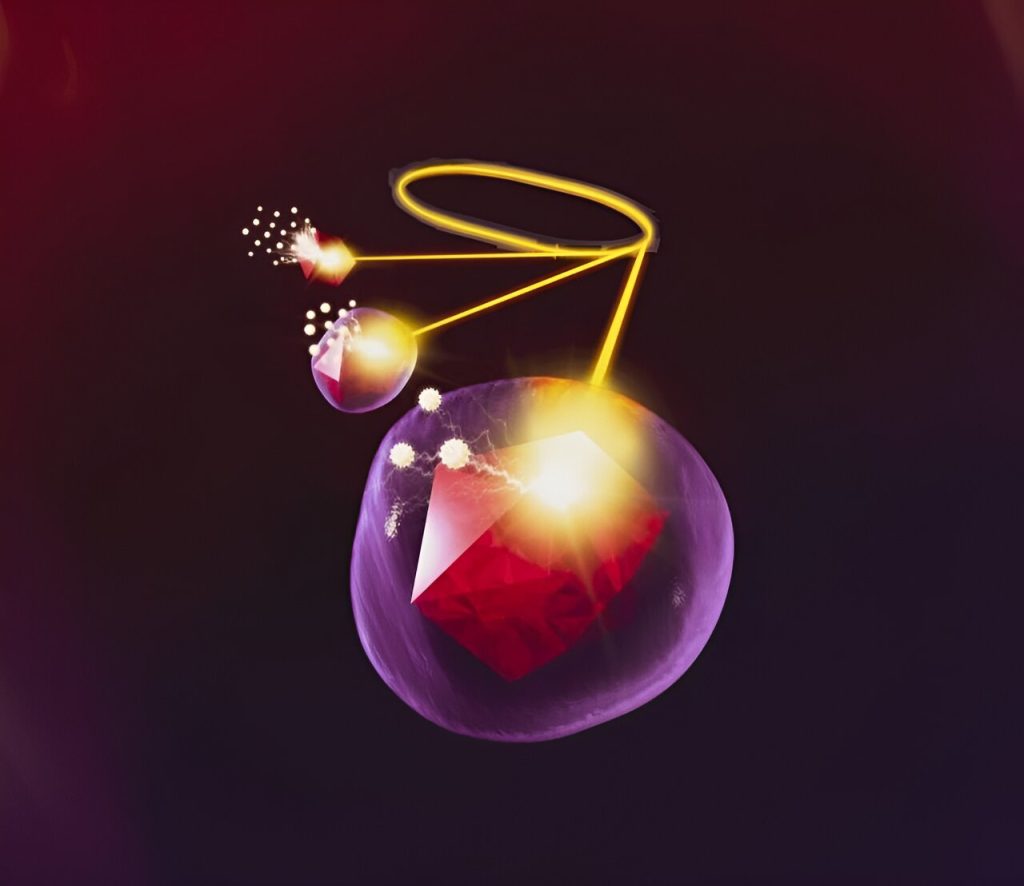
SSRL’s X-ray facilities and transition edge sensor reveal information about the nanodiamond hidden below a silica coating. Irradiated electrons escape from the nanodiamond’s surface, travel through the silica and are collected as signals. The thicker the coating, the fewer electrons make it to the surface. Understanding the chemistry of silica coatings will help researchers optimize silica shells and try other materials as coatings, expanding nanodiamonds’ applications in quantum computing and biolabeling. Credit: Greg Stewart/SLAC National Accelerator Laboratory
Coating something rare—tiny shards of diamond—with the main ingredient in sand might sound unusual, but the end result turns out to have a number of valuable applications. The trick is, nobody knows for sure how the two materials bond.
Now, researchers from San Jose State University (SJSU) report in the journal ACS Nanoscience Au that alcohol chemical groups on a diamond’s surface are responsible for usefully uniform silica shells, a result that could help them to create better silica-coated nanodiamonds—tiny tools with applications from biolabeling of cancer cells to quantum sensing.
The team unraveled the bonding mechanism thanks to powerful X-rays generated by the Stanford Synchrotron Radiation Lightsource (SSRL) at the DOE’s SLAC National Accelerator Laboratory.
“Now that we know these finer details—how the bond works instead of just guessing—we can better explore new diamond hybrid systems,” said Abraham Wolcott, the study’s principal investigator and an SJSU professor.
Much of Wolcott’s work concerns nanodiamonds, synthetic diamonds shattered into pieces so tiny that you’d need 40,000 of them to span the width of a single human hair. Theoretically, nanodiamonds have perfect carbon lattices, but occasionally a nitrogen atom sneaks in and replaces a carbon atom next to a missing carbon atom. It’s technically a defect, but it’s useful—the defect responds to magnetic fields, electric fields and light, all at room temperature, meaning nanodiamonds have many applications.
They can be used as qubits, the basic unit for a quantum computer. Hit them with green light, and they glow red, so biologists can put them in living cells and track them as they move. But scientists can’t easily program nanodiamonds to go where they want, and diamond edges are pointy and can rupture cell membranes.
Coating them with silica solves both problems. Silica forms a smooth, uniform shell that covers the sharp edges. It also creates a modifiable surface, which scientists can decorate with tags to direct the particles toward specific cells, like cancer cells or neurons. “The diamond with silica shell becomes a controllable system,” Wolcott said.
But for some time, Wolcott said, scientists have disagreed on how that shell forms. His team showed that ammonium hydroxide with ethanol, chemicals normally included in the coating process, produces many alcohol groups on the nanodiamond surface, and those alcohols facilitate the growth of the shell.
“Nobody was able to explain it for over 10 years,” Wolcott said, “but we were able to tease out that information.”
After studying the particles with transmission electron microscopes at the DOE’s Lawrence Berkeley National Laboratory Molecular Foundry, the researchers shot SSRL X-rays at nanodiamonds to explore the surfaces hidden below the silica coating.
SSRL’s transition edge sensor—a super-sensitive thermometer that collects temperature changes and converts them to X-ray energies—revealed which chemical groups were present on the nanodiamonds’ surfaces.
Using a second technique—X-ray absorption spectroscopy (XAS)—the team generated mobile electrons on the nanodiamond surface, then caught them as they traveled through the silica shell and escaped. The thicker the coating, the fewer electrons made it to the surface. The signals acted like a tiny measuring tape, showing the thickness of the silica coating on the nanometer scale.
“XAS is powerful because you can detect something that is submerged, that’s hidden—like diamond underneath a silica shell,” Wolcott said. “Folks have never done this with nanodiamonds before, so in addition to figuring out the bonding mechanism, we’ve also shown that XAS is useful for material scientists and chemists.”
In the future, Wolcott, who is known for providing hands-on research opportunities, wants to put students to work coating nanodiamonds with other materials. Titanium, zinc and other metal oxides, for example, could open new avenues in quantum sensing and biological labeling applications.
“Nanodiamonds are incredible microtools with immediate applications,” said Karen Lopez, a biomedical engineering Ph.D. student at the University of California, Irvine who, like the other SJSU authors, worked on the study as an undergraduate. “Now that we understand how the silica shell forms, we can begin optimizing it and expanding to other types of materials.”
More information: Perla J. Sandoval et al, Quantum Diamonds at the Beach: Chemical Insights into Silica Growth on Nanoscale Diamond using Multimodal Characterization and Simulation, ACS Nanoscience Au (2023). DOI: 10.1021/acsnanoscienceau.3c00033
Provided by SLAC National Accelerator Laboratory
Citation: Scientists unravel the chemical mechanism behind silica-coated nanodiamonds (2023, September 20) retrieved 24 September 2023 from https://phys.org/news/2023-09-scientists-unravel-chemical-mechanism-silica-coated.html
This document is subject to copyright. Apart from any fair dealing for the purpose of private study or research, no part may be reproduced without the written permission. The content is provided for information purposes only.

